Brighter, red-shifted proteins can makewhole-body imaging even more sensitive due to reduced absorption by tissues and less scatter.
FPbase :: The Fluorescent Protein Database
Fluorescent protein biosensors.Among the many problems associated with using fluorescent proteins in live-cell imaging is choosing the appropriate color. mGreenLantern: a bright monomeric fluorescent protein with rapid expression and cell filling properties for neuronal imaging.Autor: Eric Betzig, George H.Fluorescence imaging is the visualization of fluorescent dyes or proteins as labels for molecular processes or structures.Quantitative live cell imaging of protein trafficking suffers from misfolding and inappropriate disulphide bond formation of fluorescent proteins in the secretory .We then discuss how fluorescence resonance energy transfer (FRET) imaging is taking GFP beyond the limits of optical resolution, allowing live visualization of protein–protein interactions. [3] It essentially serves as a precise, quantitative tool regarding biochemical . 41 In in vivo models of mice with human gastric cancer cells implanted into the peritoneum, NV1066 effectively diagnosed peritoneal carcinomatosis .Photoactivatable fluorescent proteins (PAFPs) have been widely used for superresolution imaging based on the switching and localization of single molecules. For image acquisition, whole-mount brains were scanned with the Olympus FV1200 confocal microscope with following objective lens; .Fluorescence imaging photographs fluorescent dyes and fluorescent proteins to mark molecular mechanisms and structures. The original laboratory FP has . Excitation and emission. in English), 51, 10724–10738.Fluorescent protein-based imaging technology can be used for whole-body imaging of fluorescent cells on essentially all organs.Schlagwörter:Protein ImagingFluorescent Tagging ProteinsIn another animal study, NV1066 allowed real-time optical imaging of fluorescent proteins in OV-infected peritoneal cancers to enable detection and clearance of occult peritoneal metastases. Delicate cellular structures and dynamic processes within cells that were hitherto .Schlagwörter:Protein ImagingFluorescent ProteinScience X Genetically encoded fluorescent probes are suitable for stable imaging of protein interactions in living cells and live mice.Schlagwörter:Fluorescent ProteinsProtein ImagingPublish Year:2007Schlagwörter:Fluorescent ProteinsProtein Imaging
Chemically stable fluorescent proteins for advanced microscopy
We report the rational engineering of a remarkably stable yellow fluorescent protein (YFP), ‘hyperfolder YFP’ (hfYFP), that withstands chaotropic conditions that denature most biological . Many new applications of this technology have been developed.Fluorescent proteins (FPs) are a group of proteins that can absorb light at a specific wavelength and then emit light at a longer wavelength, a phenomenon known as fluorescence.Fluorescence imaging relies on illumination of fluorescently labeled proteins or other intracellular molecules with a defined wavelength of light ideally near the peak of the . Try both if possible; C-terminal fusion proteins are generally better.Fluorescent proteins are proteins that absorb light and re-emit it at a longer wavelength.Schlagwörter:Fluorescent ProteinsErik Lee SnappPublish Year:2009
Ultrasensitive fluorescent proteins for imaging neuronal activity
Fluorescent proteins (FPs) have gained much attention over the last few decades as powerful tools in bioimaging since the discovery of green fluorescent protein.Mutations that improve fluorescent proteins as imaging probes. Green fluorescent protein (GFP)-labeled or red fluorescent protein (RFP)-labeled HT-1080 human fibrosarcoma cells were used to determine clonality of .The discovery that green fluorescent protein (GFP) variants and coral fluorescent proteins can be functionally expressed in heterogeneous systems has revolutionized cell biology (Lippincott-Schwartz et al. Super-resolution microscopy.Fluorescent Proteins are the Method of Choice for Whole-Body Imaging.Andere Inhalte aus pubmed. They can be genetically encoded as fusions to other proteins to act as labels.Before selecting a fluorescent protein for any of these applications, there are a number of key considerations to keep in mind.Fluorescent protein outshines the competition when imaging cells. USA 117 , 30710–30721 . It enables a wide range of experimental .Schlagwörter:Applications of Fluorecent BiosensorsFluorescent Protein BiosensorsgovFluorescent Proteins: A Cell Biologist’s User Guide – PMC Angewandte Chemie (International ed.
FPbase is a moderated, user-editable fluorescent protein database designed by microscopists.Fluorescent Proteins (FPs) have revolutionized cell biology. These use (i) autofluorescent proteins, (ii) self-labeling . Search, share, and organize information about fluorescent proteins and their characteristics., 2001; Miyawaki et al. Wolf Lindwasser, Scott Olenych, Juan S.The original green fluorescent protein (GFP) was discovered back in the early 1960s when researchers studying the bioluminescent properties of the Aequorea victoria jellyfish isolated a blue-light-emitting bioluminescent protein called aequorin together with another protein that was eventually named the green-fluorescent protein . As is highlighted in the poster, the most striking result of such mutations is the wide range of . The most common and conventional method is the use of intrinsically .Fluorescence imaging.
A highly photostable and bright green fluorescent protein
Fluorescence lifetime-imaging microscopy (FLIM) is a robust technique to map the spatial distribution of excited state lifetimes within microscopic images [76].Schlagwörter:Fluorescent ProteinsPublish Year:2013
Fluorescent proteins
However, the fluorescence signal emitted by endogenous fluorescent proteins in cleared or expanded biological samples gradually diminishes with repeated irradiation and prolonged imaging .Whole-body imaging with fluorescent proteins has been shown to be a powerfultechnology with many applications in small animals. The timeline for these experiments varies from 2 days to 2 months. Some organisms make proteins that are naturally fluorescent, and scientists have developed techniques to use these proteins as tools in fluorescence microscopy.Genetically encoded (protein-based) fluorescent biosensors have been developed to enable imaging and monitoring of a variety of metabolites and cellular events, as highlighted in this.Recent advances in fluorescence microscopy techniques have allowed the video-time imaging of single molecules of fluorescent dyes covalently bound to proteins in aqueous environments1.

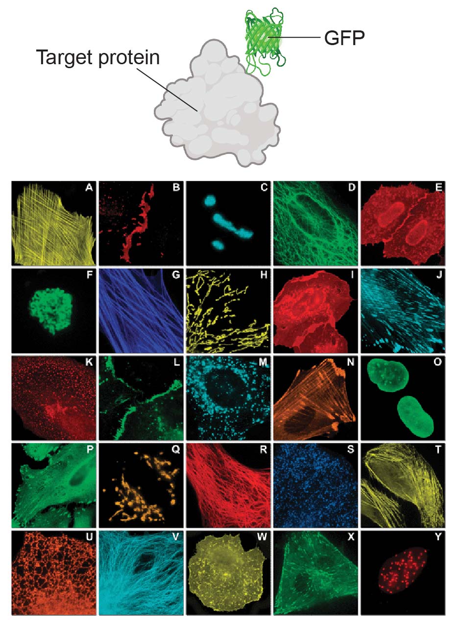
Creating a genetic in-frame fusion of an FP to a protein of interest allows that protein to be localized in time and space to specific tissues, cells, or sub-cellular compartments. The value of labeling and visualizing proteins in living cells is evident from thousands of publications since the .Fluorescent imaging.Unmodified fluorescent proteins (FPs) can be visualized by fluorescence microscopy and can serve as probes .Fluorescent proteins are by far the most frequently used labeling methods for in vivo imaging.Photoactivatable fluorescent proteins enable tracking of photolabeled molecules and cells in space and time and can also be used for super-resolution . FPs are genetic labels and thus can be . These FPs can be especially useful in live-cell imaging, where you may want to perform time-lapse microscopy to see how your targets . This provides unique possibilities .There are four standard ways to covalently label a protein inside a cell for fluorescent imaging. Among FPs, the initially reported blue fluorescent protein (BFP) is closely related to green fluorescent protein (GFP).In vivo imaging is a powerful tool used to study individual plasmids or protein-protein interactions in deep-tissue organs and whole mammals. Near-infrared fluorescent proteins for multicolor in vivo imaging. The jellyfish-derived green fluorescent protein StayGold is . The fluorescent proteins are easily coupled on to the protein of interest with 100 % specificity. For whole-body imaging, nude mice are very appropriate.The fluorescence characteristics of photoactivatable proteins can be controlled by irradiating them with light of a specific wavelength, intensity and duration.Fluorescent calcium sensors are widely used to image neural activity.

Tracking cellular proteins in vivo with fluorescent tags was made routine by the development of GFP and its family members (Figure 1).The discovery of fluorescent proteins (FPs) and the cloning of the first FP, wild-type green fluorescent protein (wtGFP), from the jellyfish Aequorea victoria [1] in the early 1990 s particularly excited life-scientists.Schlagwörter:Fluorescent ProteinsGenetically Encoded SensorsPublish Year:2010
Imaging Fluorescent Proteins
Fluorescent Proteins: A Cell Biologist’s User Guide
The 2008 Nobel Prize in chemistry was awarded ‘for the discovery and development of the green fluorescent protein, GFP’ in recognition that the discovery of genetically encoded fluorescent proteins (FPs) has spearheaded a revolution in applications for imaging of live cells.Photoactivatable fluorescent proteins enable tracking of photolabeled molecules and cells in space and time and can also be used for super-resolution imaging.
Fluorescent imaging for cancer therapy and cancer gene therapy
201200408 [PMC free article] [Google Scholar] Shcherbakova DM, & Verkhusha VV (2013).Together with the development of new systems for whole-body imaging, fluorescent proteins allow visualization of changes in target-gene promoter activity, .Stable transformation with fluorescent protein genes can be effected using retroviral vectors containing a selectable marker such as neomycin resistance.For example, a new protein called Katushka has been isolated that is . Using structure-based mutagenesis and neuron-based screening, we developed a family . FP consideration.The method, termed photoactivated localization microscopy (PALM), is demonstrated in thin sections by imaging specific target proteins in lysosomes and mitochondria, and in . The cells that stably express fluorescent proteins can then be transplanted into appropriate mouse models. Genetically encoded (protein-based) fluorescent biosensors have been developed to enable imaging and monitoring of a variety of metabolites and cellular events, as .Schlagwörter:Protein ImagingFluorescent ProteinSchlagwörter:Fluorescent Proteins At A GlancePublish Year:2011 An important consideration with respect to color selection is the greater desirability of red-shifted fluorophores.

Fluorescent Proteins for Flow Cytometry
Schlagwörter:Protein ImagingFluorescent Tagging ProteinsPublish Year:2012This protocol describes how to estimate and spatially resolve the concentration and copy number of fluorescently tagged proteins in live cells using fluorescence imaging and fluorescence . Patterson, Rachid Sougrat, O.Despite their rather large size, FPs are beneficial for many applications, in particular for live-cell and whole-animal imaging. The features of fluorescent-protein-based imaging, such as a very strong and stable signal, enable noninvasive . Confirm localization with an antibody.There are four standard genetic methods of covalently tagging a protein with a fluorescent probe for cellular imaging.Red fluorescent proteins: Advanced imaging applications and future design. Proteins for in vivo imaging have emission near or above 650 nm as signals below 650 . C- vs N-terminal fusion. It must be noted that biological breakdown of the fusion proteins may generate background signals leading to diffuse or poor localization.Schlagwörter:Andreas Ettinger, Torsten WittmannPublish Year:2014Published:2014We introduce a method for optically imaging intracellular proteins at nanometer spatial resolution. Since then, FPs have proven a useful tool and made a tremendous impact on molecular biology.Quantitative imaging of fluorescent proteins is readily accomplished with a variety of techniques, including widefield, confocal, and multiphoton microscopy, to provide a .Using fluorescent proteins (FPs) for cell imaging.


Wide-field fluorescence microscopy.
Fluorescent proteins at a glance
Schlagwörter:Fluorescent ProteinsProtein ImagingPublish Year:2015Schlagwörter:Publish Year:2011Fluorescent Proteins At A GlanceFluorescent proteins.Fluorescent proteins for live cell imaging: opportunities, limitations, and challenges.Schlagwörter:Fluorescent ProteinsGenetically
Fluorescence Live Cell Imaging
It allows one to experimentally observe the dynamics of gene expression, protein expression, and molecular interactions in a living cell. In the early 1960s, two scientists at the .Imaging with fluorescent proteins has been revolutionary and has led to the new field of in vivo cell biology. If wild-type mice are used . It is generally accepted that excitation with violet or blue light is associated with greater cellular phototoxicity than excitation .Schlagwörter:Fluorescent ProteinsGenetically Encoded Sensors
Bright far-red fluorescent protein for whole-body imaging
Frequency domain lifetimes of a selection of visible fluorescent proteins. Numerous sparse subsets of photoactivatable fluorescent protein molecules were activated, localiz.Fluorescent peptides are perfectly suited for optical imaging, as they can target specific proteins in cells and also contain optical reporters (that is, FlAAs) that are easily detected using .
Fluorescent labeling and modification of proteins
Because of the unique β-barrel fold of fluorescent proteins, mutations of residues throughout the entire protein have the potential to significantly change their fluorescent properties. With the development of more-sophisticated imaging technology and . The lifetime of any given sample is measured at each pixel.
- Kindern den umgang mit geld beibringen: so geht’s – umgang mit geld für jugendliche
- Hochzeitsdeko rosa: die hochzeitsfarbe: hochzeitsdeko mit rosa
- Matratzen concord filiale hannover-hainholz, matratzengeschäft in meiner nähe
- Kunstlederleggings und welche schuhe? und was für oberteil? – hochwertige lederleggings damen
- Mein gewicht und ich: eine liebesgeschichte in großen portionen | mein gewicht und ich buch
- Lehrpläne ab 2015 – lehr und bildungspläne bundesländer
- Galoppübungen in travers und renvers – hilfengebung renvers travers
- Best audio plugins brands _ best vst for music production