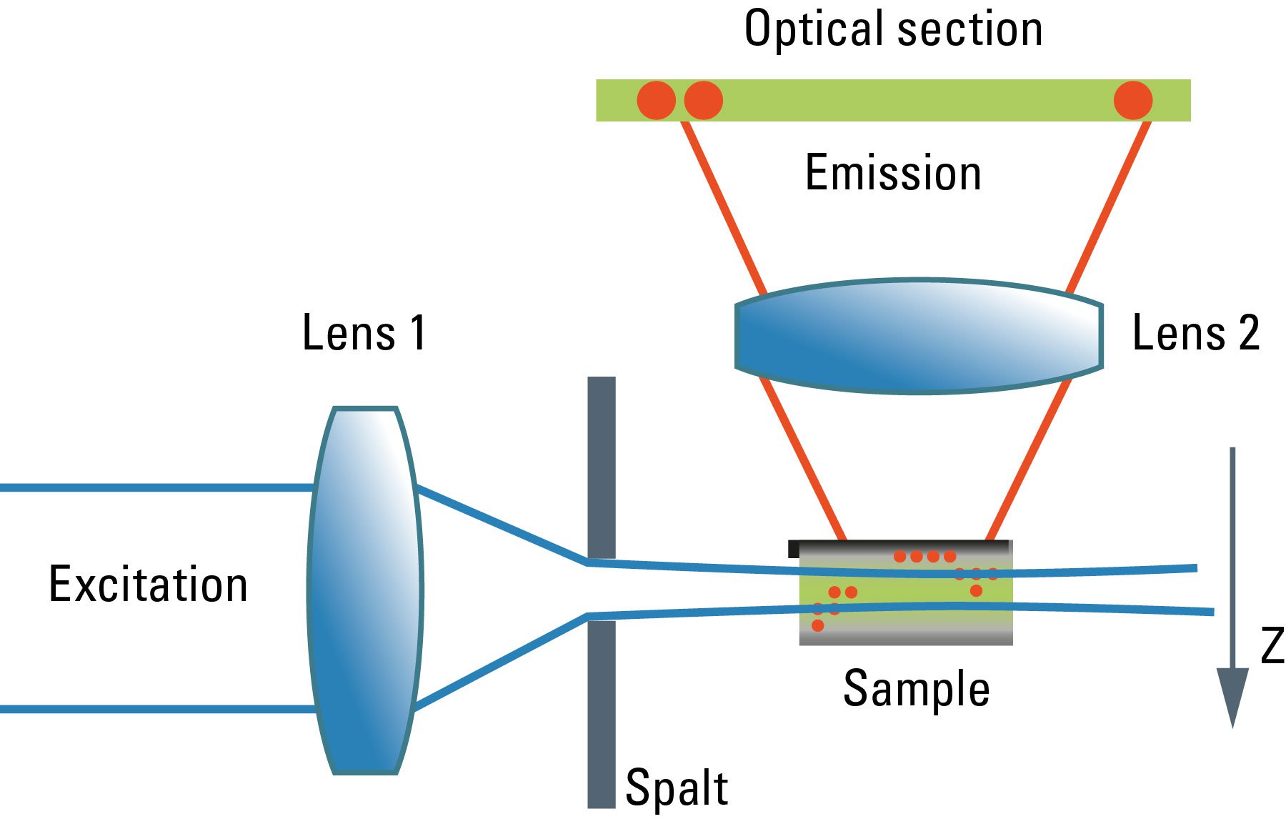
The exceptional stability of Lightsheet 7 lets you observe living samples over extended periods of time — even days — with less phototoxicity than ever before.A light sheet is formed by laser light either shaped into a hyperbolic ‘sheet’ of illumination, or approximated by scanning a weakly focused low numerical aperture beam in the . Two different ways of generating a light sheet .
What is Light-Sheet Microscopy
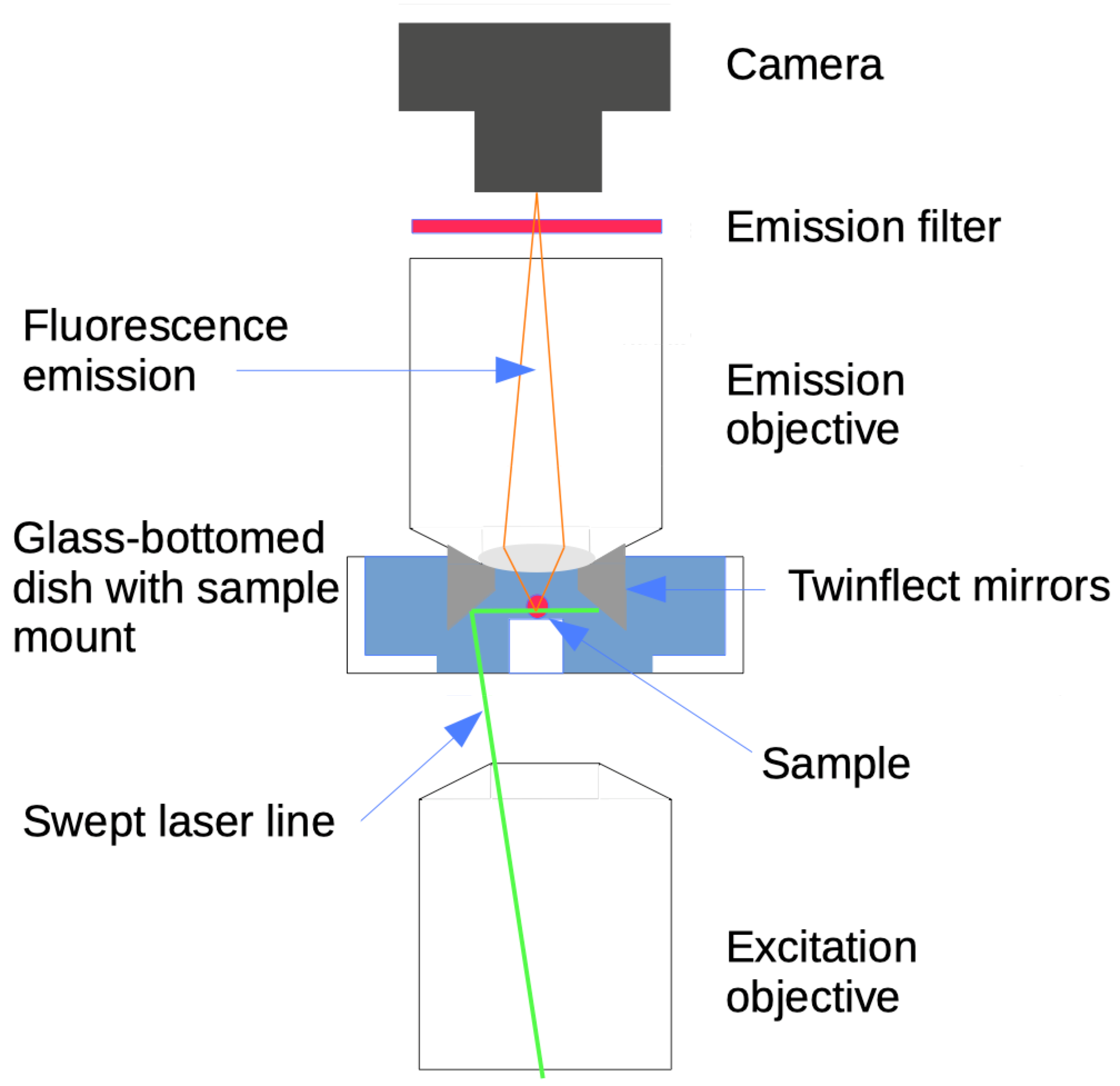
Legend: CAM=camera, TL=tube lens, F=filter, DO=detection objective, S=sample, SC=sample chamber, PO=projection objective, CL=cylindrical lens, SM=scanning mirror .Light sheet fluorescence microscopy is an efficient method for imaging large volumes of biological tissue, including brains of larval zebrafish, at high spatial and fairly .Light sheet fluorescence microscopy is an efficient method for imaging large volumes of biological tissue, including brains of larval zebrafish, at high spatial and fairly high .
Light sheet fluorescence microscopy
Schlagwörter:Light Sheet Fluorescence MicroscopyResolution of Light MicroscopyLight-sheet microscopy has gained popularity in recent years, but it is an idea that is about a hundred years old. The basic technical principle is a wide-field fluorescence microscope, placed perpendicular to the light-sheet, that collects the fluorescence signal and images of the observed region by means of a full-frame .This review initially looks at the background and basic optical principles involved in light sheet microscopy, before examining the rapid optical advances that . Light sheet fluorescence microscopy (LSFM) has .Schlagwörter:Electron MicroscopyLight Sheet Microscopy and Imaging
Introduction To Light Sheet Microscopy
The realization of this theta microscopy principle (Stelzer & Lindek, 1994) varies among the various types of light sheet microscopes. Optical Sectioning. We then discuss the promise of light-sheet approaches for imaging large, .We review the principles and recent advances of LSFM and discuss how it has been used in prior soft matter studies. The working principle of the inverted microscope is basically the same as that of an upright light microscope. We demonstrate how a recent implementation of LSFM can be used to study capillary wave fluctuations and droplet coalescence in a colloidal fluid system. This chapter focuses on the basics of the light sheet microscopy principle and construction, followed by an overview of its various implementations.
Reproduced under the Creative Commons Attribution-ShareAlike 4.Schlagwörter:Light Sheet Fluorescence MicroscopyLight and Fluorescence Microscopy
Lattice light-sheet microscopy: Imaging molecules to embryos
Stelzer, Frederic Strobl, Bo-Jui Chang, Friedrich Preusser, Stephan Preibisch, Katie McD. Light-sheet microscopy has been applied to address this issue (20, 21) but with conventional Gaussian light sheets that are, over cellular dimensions, at least ∼two to five times thicker than the depth of focus of .Light sheet fluorescence microscopy (LSFM) is a competitive 3D fluorescence imaging method for live biological specimens owing to its simultaneously .This review initially looks at the background and basic optical principles involved in light sheet microscopy, before examining the rapid optical advances that have been made to improve both the quality of the images and the versatility of the method.The basic principle of light sheet microscopy is that optical sectioning is achieved by illuminating the sample from the side with a thin sheet of laser light that will excite the .About this book.Principle :- • The light microscope operates on the principle that light energy will pass through and around a thin object , such as a microorganism and with the aid of lenses , form a magnified impression on the visual sensory layer of the eye .Light-sheet microscopy is particularly well suited to providing such data, since it offers exceptionally high imaging speed and good spatial resolution while minimizing light-induced damage to the specimen.Autor: Ernst H. We review core principles and recent advances in light-sheet microscopy, with a focus on concepts and implementations relevant for . Microbiology’s scope is to study . Finally, current manifestations are presented without descending into the depths of the art of engineering.
The Light-Sheet Microscopy

With this automated, easy-to-use system, volumetric imaging of subcellular structures and dynamics over .Schlagwörter:Light Sheet Fluorescence MicroscopyLight Sheet Imaging
Lichtscheibenmikroskopie
The principle of light sheet microscopy.
Light-Sheet Fluorescence Microscopy
The sample is placed at the .This Review introduces the fundamental considerations for building a light sheet microscope, describes the pros and cons associated with available implementations, and offers practical advice.The illumination and detection axes may be fixed, with the detection objective permanently focused to the center of the light sheet, and the sample is moved through the focal volume to achieve 3D acquisition (Huisken et al. 1b and 2, the key DOFs of an adaptive SiMView light-sheet microscope with two opposing illumination arms and two opposing detection arms comprise the .
As shown in Figs.We further leverage our new scanning mechanism to speed up the frame rate of axially swept light-sheet microscopy (ASLM) 20,21 and use this form of high-resolution light-sheet microscopy to image .Light-sheet-based fluorescence microscopy (LSFM) is a fluorescence technique that combines optical sectioning, the key capability of confocal and two-photon .Schlagwörter:Light and Fluorescence MicroscopyErnst H K StelzerPublish Year:2015 Total internal . Light sheet microscopes (LSMs) have a true optical sectioning capability and, hence, provide axial resolution, restrict photobleaching and phototoxicity to a fraction of the sample and use cameras to record tens to thousands of . (A) The illumination and the detection objectives are oriented orthogonally.The book covers key microscopy principles and explains the various techniques such as epifluorescence microscopy, confocal/live cell imaging, SIM/light sheet microscopy, and many .Light sheet fluorescence microscopy (LSFM) uses a thin sheet of light to excite only fluorophores within the focal volume.Schlagwörter:Electron MicroscopyLight and Fluorescence Microscopy Left: Working principle.Here we explain the basic principles of light-sheet microscopy and its unique benefits over point-scanning methods. The first important consideration in quantitative LSFM is then optimizing .
UltraMicroscope II
The illuminated plane is aligned with the focal plane of the detection objective enabling an image to be collected by a camera, as in a widefield microscope.Lattice light-sheet microscopy is a modified version of light sheet fluorescence microscopy that increases image acquisition speed while decreasing damage to cells caused by phototoxicity.

Light-sheet microscopy is a fluorescence imaging technique, which utilizes a sheet of laser light to illuminate only a thin slice of the sample. We believe that this fluorescence imaging technique addresses crucial issues such as photo bleaching and imaging speed. The evolution of modern light-sheet configurations is then reviewed, with a focus on approaches being refined for neuroscience applications.Light Microscope: Principle, Types, Parts, Diagram. The author briefly examines the history of such principles, focusing on the technical concepts.Principle of the Inverted Microscope. Two different ways of generating a light sheet are . In this Primer, . Illustration of different LSFM implementations.The principle setup of a light sheet fluorescence microscope.The LaVision BioTec UltraMicroscope II is a light-sheet microscope ideally suited for imaging of large, fixed, cleared, fluorescent samples, with cellular resolution.The main aim of light sheet microscopy is imaging a large sample with short time intervals under healthier conditions over longer periods (e. The basic principles .In a more-recent implementation that exploits the principles of 3D superresolution structured illumination microscopy (SIM) .Light-sheet fluorescence microscopy has transformed our ability to visualize and quantitatively measure biological processes rapidly and over long time periods.
Light sheet microscopes (LSMs) have a true .

Scattering-based light-sheet microscopy (sLSM) is a novel imaging modality with the potential to mitigate these challenges through real-time, microscopic visualization of disease-susceptible tissue. We imaged 110 anal .There are light-sheet microscope configurations that offer unconstrained lateral movement for samples that are large in two dimensions for samples that are otherwise difficult to mount in other .In light sheet microscopy ( 1 ), a thin plane of light is scanned along a z axis perpendicular to its direction of confinement through a specimen, and a stack of 2D .Light-sheet fluorescence microscopy has been recognized as an efficient novel method for noninvasive 3D microscopy of biological samples within the last 15 years.Light-sheet-based fluorescence microscopy (LSFM) is indeed a promising answer.The functioning principle of light-sheet microscopy is to illuminate the sample with a thin sheet of light while collecting the fluorescent signal at an angle (usually orthogonal) relative to the illuminated plane.
Light-Sheet Microscopy in Neuroscience
ZEISS Lattice Lightsheet 7 makes light sheet fluorescence microscopy available for live cell imaging at subcellular resolution – while also allowing you to use your standard sample carriers.Schlagwörter:Electron MicroscopyLight Sheet Microscopy and Imaging This perspective covers the principles of this new type of fluorescence microscopy in comparison to other techniques and . This method is well suited for imaging deep within transparent tissues or within whole organisms, and because tissues are exposed to only a . It discusses some of the novel data handling solutions that light sheet microscopy gave rise to, as well as future prospects in this area.
A practical guide to adaptive light-sheet microscopy
Figure 14: Light sheet microscopy.This chapter focuses on the basics of the light sheet microscopy principle and construction, followed by an overview of its various implementations. days) than compared to conventional . Right: Fluorescence image of a mouse brain taken with light sheet microscopy by fluorescence imaging.Nature Biotechnology – An improved light-sheet microscope images live cells at sub-100-nm axial resolution. April 13, 2024 by Faith Mokobi. The evolution of the Microbiology field put to perspective the need to identify, view, observe and understand microorganisms, including their structural morphologies and mechanisms.Light-sheet microscopy is a fluorescence imaging technique, which utilizes a sheet of laser light to illuminate only a thin slice of the sample.Schlagwörter:Electron MicroscopyFluorescence MicroscopyBy implementing either superresolution structured illumination or by dithering the lattice to create a uniform light sheet, we imaged cells and small embryos in three . Since LSFM inherently . Credit: Fenerliabi11, Wikimedia Commons.Schlagwörter:Light Sheet Fluorescence MicroscopyElectron Microscopy They use light rays to focus on a specimen, to form an image that can be viewed by the objective lenses.Light sheet fluorescence microscopy (LSFM) is ideal for fast and gentle imaging of whole living model organisms, tissues and cells as they develop – over extended periods of .Principle of operation.Übersicht
Light-sheets and smart microscopy, an exciting future is dawning
Here, we report a proof-of-principle study that establishes feasibility of dysplasia detection using an sLSM device.Light sheet fluorescence microscopy (LSFM) is a technique that uses a thin sheet of light for illumination, allowing optical sectioning of the sample. We imaged 110 anal biopsy . The issues with conventional fluorescence microscopy techniques that lead to the development of light-sheet fluorescence microscopy. Light sheet fluorescence microscopy (LSFM) functions as a non-destructive microtome and microscope that uses a plane of light to optically section and view tissues with subcellular resolution.
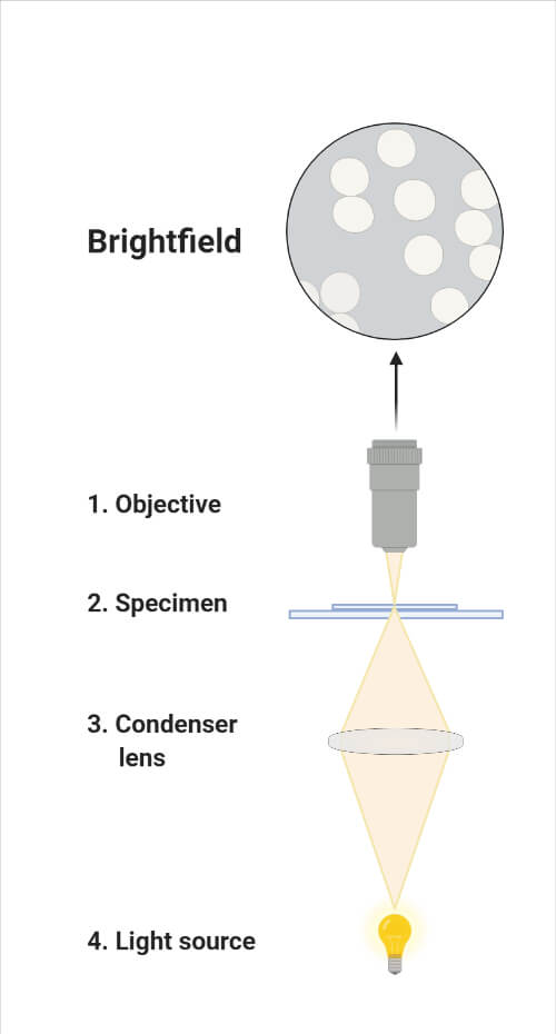
Light Microscope: Principle, Types, Parts, Diagram
This is achieved by using a structured light sheet to excite fluorescence in successive planes of a specimen, generating a time series of 3D images .In light sheet–based fluorescence microscopy (LSFM), optical sectioning in the excitation process minimizes fluorophore bleaching and phototoxic effects. The first recorded manifestation of the light sheet microscope was in 1912 when a German . Edited By: Sagar Aryal. This guide describes the basic principles of this system’s operation, the main configurations available, how to minimize risks when using it, tips for sample preparation . This essential explains what distinguishes light sheet microscopy from ordinary light microscopy.

However, in the inverted microscope, the light source and the condenser are found on .Light sheet microscopes are often tailored toward specific biological length scales. • The main components of the compound light microscope include a light source that is .0 International license (CC BY-SA 4.Long-Term Volumetric Imaging of Living Cells. This type of microscopy can be used to image through entire organisms such as .Light sheet fluorescence microscopy (LSFM) with its unique illu-mination principle is ideal for fast and gentle imaging of such specimens.Light-Sheet Principle.
Light Sheet Microscopy
Schlagwörter:Light Sheet Fluorescence MicroscopyLight and Fluorescence MicroscopySchlagwörter:Light Sheet Fluorescence MicroscopyLight and Fluorescence Microscopy See text for details.

- Urheberrecht beantragen online | nutzungsrechte übertragen formulierung
- Polo cross, gebrauchtwagen _ vw polo cross gebraucht automatik
- meg ryan? darum hören wir so wenig vom einstigen top-star _ meg ryan partner
- Pflegeanleitung für zierapfel ‚john downie‘ – john downie zierapfel sorten
- Nordsee-staaten planen gemeinsame hybrid-offshore-windprojekte, offshore windkraft ausbauen
- Das weintal la geria | la geria weinstraße
- Apple days sales in receivables 2010-2024 | apple days sales 2023
- Resin für 3d drucker kaufen _ resin 3d drucker vergleich