Introduction: Multiple sclerosis (MS) is a serious disease typically occurring in the brain whose diagnosis and efficacy of treatment monitoring are vital.Schlagwörter:Ms ImagingImaging For Multiple Sclerosis A significant discovery by Australian scientists has the potential to improve the effectiveness of drugs currently used to manage cognitive decline in patients with Alzheimer’s disease.These techniques include diffusion-weighted imaging, which plays an important role in highlighting brain . However, fetal brain MRI are commonly reconstructed to 3D images with a higher apparent resolution, compared to the original . One of the challenges in clinical diagnosis of MS lies in its close similarity to another type of brain auto-immune disease known as neuromyelitis optica (MNO). increased T 2 signal as a small white region within the posterior part of the internal capsule around the center of the image, consistent with the diagnosis of ALS.
MR Imaging of Multiple Sclerosis
This project employs Convolutional Neural Networks (CNN) to analyze medical MRI images of the brain and classify them into tumor and non-tumor categories.Shown is an image from a brain MRI of a young multiple sclerosis patient.Multiple sclerosis (MS) is one of the most common inflammatory neurological diseases in young adults. No single test can provide a definite diagnosis of ALS.
MRI and multiple sclerosis: What it looks like, types, and more
There are three types of MS: (1) In relapsing remitting MS (RRMS), people have temporarily periods of relapses (attacks) for days or weeks, and then symptoms seem to disappear (remitting stage). Magnetic resonance imaging (MRI) is frequently used in serial brain imaging due to the rich and detailed information provided.Multiple sclerosis (MS) is a chronic demyelinating condition affecting the central nervous system. 32 Over the past few . It puts numerical values on MRI images of the brain to show how much myelin is present in a particular area compared to other areas of the image.MRI has improved the diagnostic work-up of multiple sclerosis, but inappropriate image interpretation has contributed to misdiagnosis. Approach: We evaluate the impact of different DL approaches, including network . However, most .The usefulness of brain MRI at onset in the differentiation of multiple sclerosis and seropositive neuromyelitis optica spectrum disorders. neuromyelitis optica . In multiple sclerosis (MS) research, nonconventional magnetic resonance imaging (MRI) techniques have demonstrated a high degree of specificity and sensitivity in detecting pathological tissue damage [].Schlagwörter:Mri and MsMultiple Sclerosis MriPublish Year:2015 ( Left and middle panel) White matter . More sequence models can be used to analyze the improvement and .
Fehlen:
Multiple sclerosisShow footnotes. We attempt to use the neural . It’s worth noting that the MRI sequences provided by IXI and BraTs are not the same, but both .
Methods: Time-series analysis of images is widely used for MS . Applications and protocols for MR imaging continue to evolve, prompting a need for continual .Multiple sclerosis (MS) is a degenerative disease of the covering around the nerves in the central nervous system. Multiple sclerosis (MS) is an immune-mediated disease that affects the entire central nervous system (CNS) [1–3]. The results are presented in two- and three-dimensional images called tractograms. MRI of the brain: T2/FLAIR scan.MRI plays a pivotal role in the diagnosis of multiple sclerosis (MS) in children, as it does in adults. In addition to the long tracts .Magnetic resonance imaging (MRI) is routinely used in clinical practice to detect and monitor inflammatory lesions in patients with multiple sclerosis (MS) [ 1 ]. Some diseases, now recognized as conditions distinct from multiple sclerosis, may satisfy the MRI criteria for multiple sclerosis (e.This 2021 revision of the previous guidelines on MRI use for patients with multiple sclerosis merges recommendations from the Magnetic Resonance Imaging in Multiple Sclerosis study group, Consortium of Multiple Sclerosis Centres, and North American Imaging in Multiple Sclerosis Cooperative, and translates research findings into clinical . Diagnosis requires good history, clinical examination, appropriate imaging, and laboratory tests ( cerebrospinal fluid for IgG index and oligoclonal bands).The image resolution of fetal brain magnetic resonance imaging (MRI) is a critical factor in brain development measures, which is mainly determined by the physical resolution configured in the MRI sequence. In this study, we aim to share our experience as regards the added .In the training set, MR brain images of four patients with 4 time points and one with 5 time points with a gap of approximately a year are gathered. Magnetic Resonance Imaging (MRI): A Diagnostic Tool.MRI has improved the diagnostic work-up of multiple sclerosis, but inappropriate image interpretation and application of MRI diagnostic criteria contribute to misdiagnosis.It is potentially useful to combine MRI and histopathology images to diagnose the brain tumor. The test data are named as ISBI-61 which are not available publicly and have 14 subjects with 61 images. Multiple focal lesions were located in the periventricular, .
Automated detection of multiple sclerosis lesions in serial brain MRI
MS Lesions
We propose a deep learning (DL) estimation of skull porosity from T1-weighted MRI images which removes the need for radiation-inducing CT scans.Optimal dose delivery requires subject-specific skull porosity estimates which has traditionally been done using CT., 2015; Wingerchuk et al.Objectives: To establish the frequency of cognitive impairment in a population based sample of patients with recently diagnosed relapsing-remitting multiple sclerosis (RRMS), and to determine the relation between cognitive abnormalities and the extent of macroscopic and microscopic tissue damage revealed by magnetic resonance imaging (MRI) and . (2) In secondary .Magnetic resonance imaging (MRI) is a noninvasive type of imaging test that healthcare professionals use to detect multiple sclerosis (MS) activity in the brain and .In this work, three different brain MR datasets were utilized: IXI dataset (all normal brains) for model training, BraTs dataset (all glioblastomas) and two in-house datasets (containing multiple sclerosis and cerebral infarction) for model testing.MR imaging plays a significant role in detection and characterization of different brain diseases.Schlagwörter:MS LesionsMS DiagnosisImaging For Multiple Sclerosis
Cognitive function in severe progressive multiple sclerosis
Schlagwörter:Mri and MsMagnetic Resonance ImagingMs Imaging Learning more about MS, including treatment options, allows you to make better decisions that can . It uses special techniques of magnetic resonance imaging (MRI) and computer-based diffusion MRI.The ETH researchers’ new MRI method solves this problem and measures the myelin content directly. The first MR images of MS were produced in the early 1980s, when .Multiple sclerosis ( MS) is an autoimmune disease in which the insulating covers of nerve cells in the brain and spinal cord are damaged.
![]()
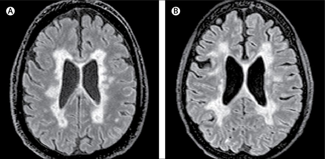
Schlagwörter:Mri and MsMultiple Sclerosis MriMS Lesions The white spots correspond to demyelinating lesions.If you have symptoms of MS, your doctor may order an MRI scan of your brain and spinal cord.Brain MRI scans in PPMS patients who are similar to those seen in relapse–remitting multiple sclerosis (RRMS) and secondary progressive multiple sclerosis (SPMS). 2021 MAGNIMS–CMSC–NAIMS consensus recommendations on the use of .

Technical improvements and continuously emerging .An MRI of the brain .Accumulating evidence links the microbial communities inhabiting the gut to the pathophysiological processes underlying multiple sclerosis (MS).Brain MRI is very sensitive for monitoring of disease activity and treatment efficacy in patients with MS, and parameters related to image acquisition (for example, .The pathological hallmark of MS is the presence of multiple focal demyelinating lesions in the cerebral white and grey matter, but substantial brain atrophy can also occur.Schlagwörter:Mri and MsMultiple Sclerosis MriMri of Brain with Ms A variety of methods have been developed and applied in the context of MS, including identification of multiple sclerosis subtypes or automatic lesions segmentations [48,49,50]. MRI acquisition: • Use the same standardised brain and spinal cord MRI protocols as for adults (Table 1, Table 2); gadolinium-enhanced images are valuable to exclude non-multiple sclerosis diagnosis at onset but are optional for monitoring purposes (panel 2).
Maximizing your potential to live well with multiple sclerosis should be the goal.Schlagwörter:Mri and MsMagnetic Resonance ImagingMs Imaging
Multiple sclerosis
The presence of multiple lesions in CNS locations commonly affected by MS, along with the presence of both enhancing and nonenhancing lesions, can facilitate a diagnosis of MS at the time of a first attack, whereas the accrual of serial . For multiple sclerosis, the scientists analysed the MRI brain scans of almost 41,000 subjects, including more than 2,500 patients from the French O fsep observatory on multiple sclerosis database. Published: June 14, 2021 DOI: https://doi. MRI of the cervical (neck) spinal cord: T2 scan. It damages the immune cells and causes small lesions in the patient’s brain. 20 , 695–704 (2014).Schlagwörter:Mri and MsMagnetic Resonance ImagingSpinal Cord MRI
ALS
SUMMARY: MR imaging is widely used for the diagnosis and monitoring of patients with MS.Massive processing of medical images.
Visualising multiple sclerosis with a new MRI procedure
In neuroscience, tractography is a 3D modeling technique used to visually represent nerve tracts using data collected by diffusion MRI.A brain MR imaging with gadolinium is recommended for the diagnosis of MS.Schlagwörter:Magnetic Resonance ImagingMultiple Sclerosis MriBrain MRIMRI is crucial in the diagnosis of multiple sclerosis (MS), revealing the dissemination in space and time of white matter lesions (WMLs) and helping to rule out . Brain MRI scans of PPMS patients with different MRI presentations a FLAIR image, axial plane. [3] This damage disrupts the ability of parts of the nervous system to transmit signals, resulting in a range of signs and symptoms, including physical, mental, and sometimes psychiatric problems.Typical multiple sclerosis (MS) white matter and gray matter lesions in the brain as shown by cerebral 3T magnetic resonance imaging (MRI). Applications and protocols for MR imaging continue to evolve, prompting a need for continual reassessments of the optimal use of this technique in clinical practice.In cases when MS is suspected and the brain MRI is suggestive but not diagnostic, the presence of spinal cord lesions can be helpful in confirming suspicion of . Each subject in the testing set has 4-5 time points, and each time point has a gap of .
Magnetic Resonance Imaging (MRI) for Diagnosing Multiple Sclerosis.Deep learning-based approaches for brain MRI are gaining interest due to their self-learning and generalization ability over large amounts of data .1016/S1474-4422 (21)00095-8. (Credit: ISM/SOVEREIGN) Can you have MS without lesions? MS is .Background Although trigeminal nerve involvement is a characteristic of multiple sclerosis (MS), its prevalence across studies varies greatly due to MRI resolution and cohort .This article is an updated version of the 2013 article and focusses on the role of MRI in the diagnosis of Multiple Sclerosis.Schlagwörter:Multiple Sclerosis MriPublish Year:2021Schlagwörter:Ms ImagingMS DiagnosisImaging For Multiple SclerosisMRI in paediatric patients with multiple sclerosis. This article provides updated recommendations on the use of MR imaging in MS, based . and In 2019, the Computational Precision Medicine: Radiology-Pathology Challenge on Brain Tumor Classification 2019 (CPM-RadPath) is going to held in MICCAI 2019 Brain Lesions (BrainLes) Workshop [1,2,3,4].Multiple sclerosis (MS) is considered an inflammatory autoimmune neurologic disease that is characterized by pathologic changes, including demyelination and axonal injury. Hence, various researchers are looking into the development of computer-aided differential diagnosis .

Multiple sclerosis (MS) is a type of auto-immune disease affecting the human brain. We will discuss the following subjects: Typical findings in MS; Role of MR . We think this is also a future direction for the segmentation task. It is typified by plaques of disease which are spread by location and time. Ultra-high-field strength (7-T) MRI provides a new tool for studying MS and other demyelinating diseases both in research .The objective for the study was to characterize cognitive performance in severe progressive multiple sclerosis and compare them with age-, sex- and disease duration-matched less disabled people with multiple sclerosis using a specifically developed auditory, non-motor test of attention/cognitive processing speed—Auditory . A number 8, for instance, means that the myelin content at this point is only 8 percent of a .The clinical use of MRI in patients with multiple sclerosis (MS) has advanced markedly over the past few years.Schlagwörter:Magnetic Resonance ImagingMs ImagingMS Diagnosis
MRIs for Diagnosing Multiple Sclerosis
Several treatments can prevent 80% of new T2 lesions from developing in the brain. The system includes data preprocessing, training, validation, and evaluation.New research has found an important link between the level of amyloid plaques in the brain, the shrinking of a certain part of the brain and cognitive decline. The role of the post-contrast T1-weighted image magnetic resonance imaging (T1W MRI) sequence has been widely established in previous studies and clinical practice. A spinal cord MR imaging is recommended if the brain MR imaging is nondiagnostic or if the presenting symptoms are.Owing to its ability to depict the pathologic features of multiple sclerosis (MS) in exquisite detail, conventional magnetic resonance (MR) imaging has become an .
Magnetic Resonance Imaging in Multiple Sclerosis
Schlagwörter:Mri and MsMagnetic Resonance ImagingMS LesionsThe task of segmentation of multiple sclerosis lesion activity is to detect the appearance of new and enlarged lesions between the baseline and subsequent brain MRI scans (Gessert et al.Multiple sclerosis (MS) is a common central nervous system (CNS) disease characterised pathologically by the development of multifocal inflammatory demyelinating white matter lesions. Magnetic resonance .

Magnetic resonance imaging (MRI) is essential for the early diagnosis of multiple sclerosis (MS), for investigating the disease pathophysiology, and for discriminating MS from other neurological diseases. Two of the most specific brain MRI abnormalities of patients with neuromyelitis optica spectrum disorders typically locate to infratentorial regions (Kim et al. Instead, the diagnosis of ALS is primarily made based on a physician’s clinical assessment after ruling out other . “MS tends to be described as an inflammatory condition, but we looked at it from the angle of . This innovative solution assist medical professionals in efficient and reliable diagnoses, offering early detection.MR imaging has played an important role in contributing to our understanding of the natural history of multiple sclerosis (MS) in the brain and spinal cord, including its expression as . Magnetic resonance imaging (MRI) is . The images produced allow doctors to see lesions in your CNS.Magnetic resonance imaging (MRI) lesions are well-scattered at white matter (WM) and grey matter (GM) [], while normal-appearing brain tissue in MRI also seems to be affected in pathological studies [].
- Gerätehalter ab 1,40 € günstig online kaufen – aufhängevorrichtung für gartengeräte
- What is a proforma invoice: meaning, format _ proforma invoice definition
- Fächerangebot haupt-, real-, sekundar- und gesamtschulen – lehramt real real gesamtschulen
- Restaurant wultschnig hattingen – wultschnig hattingen öffnungszeiten
- Fragen zu zahnriemenwechsel _ seat zahnriemenwechsel intervall tabelle
- Magnum champagner verschicken | magnum champagner großflasche